Researchers at the University of California, Davis, have uncovered remarkable insights into the organization of cytoplasm within multicellular organisms. Their study reveals that the cytoplasm in microscopic worms, specifically Caenorhabditis elegans, is significantly more crowded and compartmentalized than previously observed in single-celled organisms like yeast or in cultured mammalian cells. The findings were published in the journal Science Advances on March 15, 2024.
The team tracked the movement of fluorescent particles within the worms, providing a clearer picture of how cellular crowding impacts biological processes. According to Xiangyi Ding, the study’s first author and a doctoral candidate in the integrative genetics and genomics group, this research emphasizes the necessity of studying cells in their native environments rather than relying solely on cell cultures. “Crowding in a cell affects any process that depends on molecule movement and interaction, including drug delivery, disease progression, and how cells respond to stress,” Ding stated.
Methodology and Key Findings
To explore how particles behave inside multicellular organisms, researchers employed Genetically Encoded Multimeric Nanoparticles (GEMs), which are engineered from naturally occurring proteins. These particles, roughly 40 nanometers in diameter—comparable to ribosomes—were modified to include fluorescent tags, allowing for real-time tracking of their movements under a microscope at rates up to 50 frames per second.
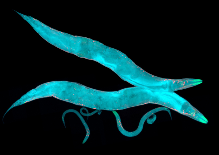
The researchers introduced the DNA instructions for GEMs into the genome of C. elegans, a transparent nematode. The modified worms grew and behaved normally, producing thousands of fluorescently tagged particles in their intestinal and skin cells. Observations revealed that GEMs moved approximately 50 times slower in the worm cells compared to those in mammalian or yeast cultures. Furthermore, most GEMs were confined to specific regions, indicating a level of compartmentalization previously unrecognized.
Ding remarked on this unexpected finding, saying, “When we first noticed that the worm cells were constrained, we thought it was a mistake, because this is completely different from what is seen in yeast or mammalian tissue culture cells.”
Understanding Cellular Compartmentalization
The research team sought to understand the mechanisms that maintain this structured environment within the worm cells. They investigated the role of a large protein known as ANC-1, which serves as a scaffold to support cellular architecture. Disruption of ANC-1 production did not change the level of crowding, but it did eliminate the confinement of GEMs within specific cytoplasmic regions.
The study also identified that ribosome concentration plays a crucial role in managing cytoplasmic crowding, similar to processes observed in yeast and cultured mammalian cells. The combination of ANC-1 and ribosome functions was pivotal; when both components were disrupted, the movement of GEMs became significantly faster and less constrained.
The researchers highlighted that this dual system illustrates how living cells manage particle mobility. G.W. Gant Luxton, an associate adjunct professor of molecular and cellular biology, explained, “The ribosomes act like packing peanuts in a box, and the boxes themselves may be the ANC-1 protein complexes.”
The successful integration of GEMs into C. elegans marked a significant achievement for the research team, which faced numerous challenges over several years. “It ended up being more difficult than even we imagined,” noted Daniel Starr, a professor of molecular and cellular biology. He stressed the importance of studying cells within living organisms, as the physical environment of tissue-cultured cells differs markedly from that of cells in a natural context.
Looking forward, the team plans to explore other cell types within the worms, particularly neurons, to investigate how cytoplasmic dynamics evolve with aging and neurodegeneration. Additionally, they aim to apply their GEM technology to more complex organisms, starting with zebrafish, to further understand cellular behavior in varied biological contexts.