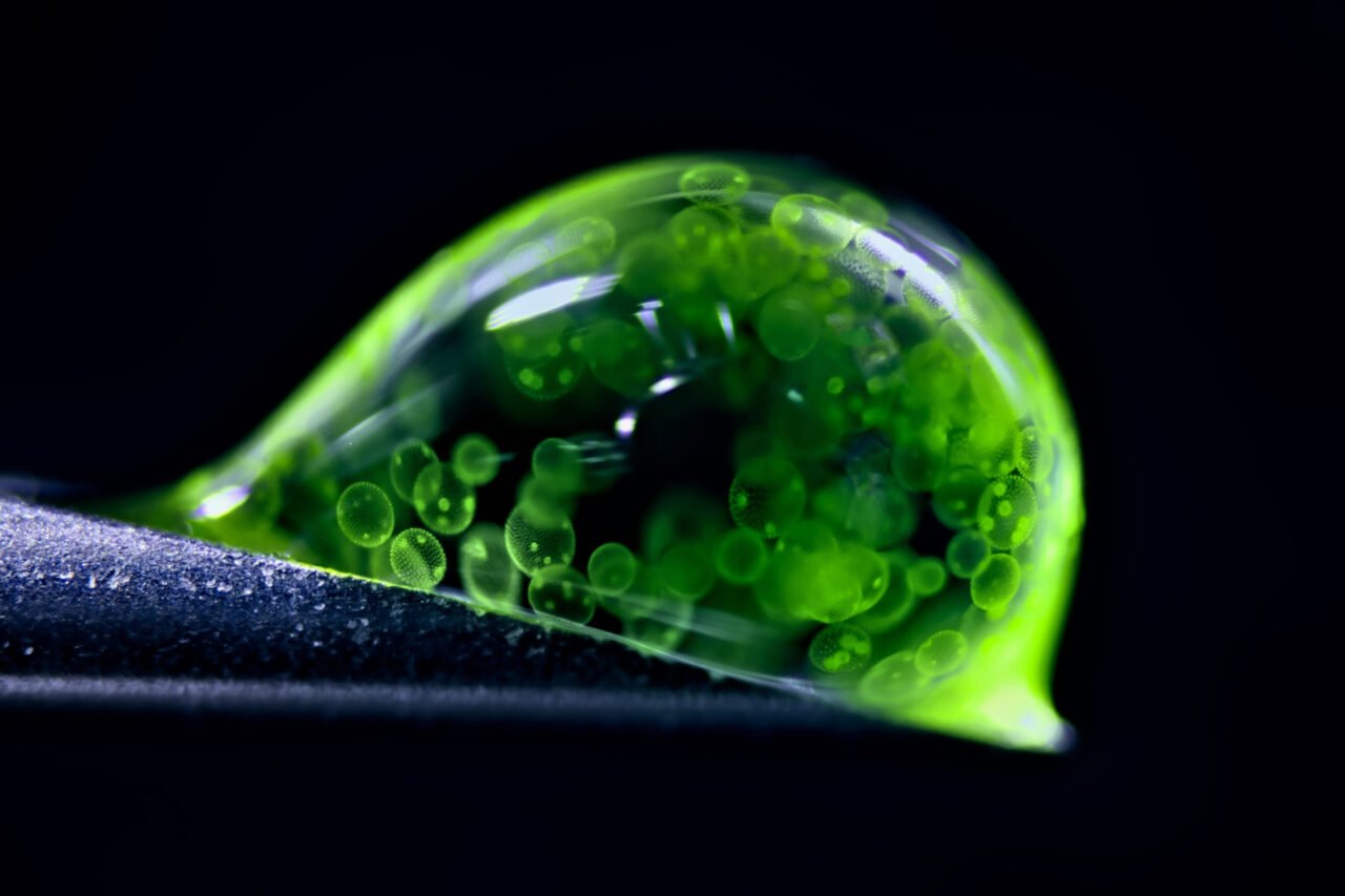
Photography has taken a fascinating turn with the results of the 2025 Photomicrography Competition, organized by Nikon. This year’s entries showcased extraordinary images that challenge our understanding of the microscopic world. Among the standout photographs is a remarkable shot by Jan Rosenboom, a chemical engineer from Germany, who captured a drop of water filled with tiny spheres of colonial algae using reflected light microscopy. His work earned him the runner-up position in the annual competition, which celebrates innovative microscopy in scientific exploration.
Each year, this competition draws contributions from professional scientists and amateur enthusiasts alike, revealing stunning snapshots of familiar objects from entirely new perspectives. Ed Cara, a judge for the 2023 competition, noted the creativity and technical skill exhibited by participants. The winners this year displayed a diverse range of subjects, from human cellular networks to the hidden worlds within unassuming mushrooms.
Highlights from the Competition
Among the notable entries was a striking image of red, diffused pigments from the fungus Talaromyces purpureogenus, captured by Wim van Egmond of the Micropolitan Museum in the Netherlands. Placing ninth in the competition, van Egmond’s photograph highlights the vibrant beauty found in microscopic life, often overlooked in everyday observations.
Another captivating entry came from researchers at the Friedrich Miescher Institute for Biomedical Research in Switzerland. Their image of a mouse colon, taken using confocal microscopy, showcased the intricate cellular structures highlighted by fluorescent probes. This technique is widely used in biomedical sciences for studying various cellular processes.
In a different vein, James Hayes of Vanderbilt University captured an insightful image of heart muscle cells, showcasing chromosomes condensing during cell division. Such images not only reveal the complexity of our bodily functions but also enhance our understanding of human biology.
Unexpected Discoveries and Natural Wonders
The competition also unveiled surprising visuals, such as an image by Stella Whittaker from the National Institutes of Health (NIH). Her photograph of induced pluripotent stem cell-derived sensory neurons resembled a swirling black hole, illustrating the fascinating complexities of cellular structures when viewed at such high magnification.
In a more alarming context, Igor Robert Siwanowicz from the Howard Hughes Medical Institute captured a striking image of marrow pollen germinating on stigma while being attacked by a filamentous fungus. This photograph starkly depicts the interdependent relationships that exist within the microscopic realm, emphasizing nature’s often brutal realities.
The competition also featured a captivating image of pollen spores suspended on a spider web, taken by John-Oliver Dum from Medienbunker Produktion in Germany. His third-place photograph, achieved through the technique of stacking multiple shots, emphasizes the delicate yet resilient structures found in nature.
Finally, the overall winner of the competition, Zhang You, an entomologist from China, captured a rare moment of a rice weevil spreading its wings on a grain of rice. This stunning photograph was a result of combining over 100 images to enhance clarity and detail, showcasing You’s dedication to ecological and insect science photography.
The 2025 Photomicrography Competition not only highlights the beauty of the microscopic world but also serves to inspire further inquiry into the intricacies of life that exist beyond our immediate perception.