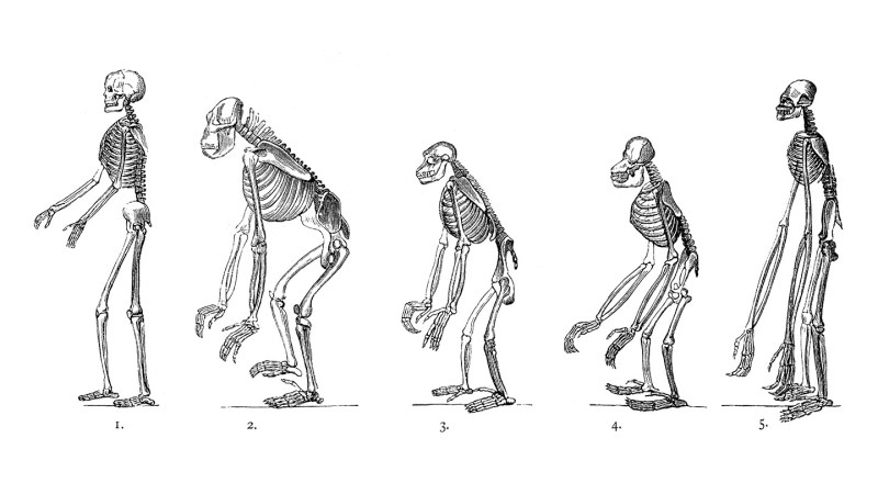
Two small genetic changes played a crucial role in the evolution of early humans, enabling them to walk upright. Researchers from Harvard University discovered that these genetic shifts significantly altered the structure of the human pelvis, transforming it from a design suited for quadrupedal movement into one that supports bipedal locomotion.
One of the changes involved a 90-degree rotation of the ilium, the uppermost part of the pelvic bone. This adjustment reoriented the muscles connected to the pelvis, facilitating a transition from climbing and running on four legs to standing and walking on two. The second genetic alteration extended the time it takes for the ilium to develop from soft cartilage into bone. These findings were published in the peer-reviewed journal Nature on September 25, 2023.
How Genetic Changes Shaped Human Anatomy
According to Gayani Senevirathne, an evolutionary biologist at Harvard, the reconfiguration of the ilium allowed muscles that previously supported movement on all fours to adapt for upright walking. Coauthor Terence Capellini emphasized that these changes were vital for redistributing the muscular system, converting it from one primarily located on the back to one that operates on the sides, thereby enhancing bipedal stability.
To arrive at these conclusions, researchers analyzed developing pelvic tissue samples from humans, chimpanzees, and mice. They employed advanced CT imaging to observe that human ilium cartilage grows horizontally rather than vertically, a significant distinction from other primates. Additionally, the transition from cartilage to bone occurs more slowly in humans, allowing for a wider, bowl-shaped pelvis essential for bipedalism.
The genetic analysis revealed that the changes corresponded to regulatory switches that control gene activity. In humans, genes responsible for cartilage formation activated in specific regions of the growing ilium, promoting horizontal growth. Conversely, bone-forming genes activated later in different locations, enabling the ilium to expand sideways rather than just thickening vertically.
Implications for Evolutionary Biology
The timing and location of these genetic changes suggest that the rewiring of developmental genes took place early in the hominid lineage, after humans diverged from chimpanzees. This research supports a core tenet of evolutionary developmental biology: significant anatomical advancements often result from subtle modifications in gene regulation rather than entirely new genes.
Anthropologist Carol Ward from the University of Missouri highlighted the importance of these findings, stating that the ability to stand on one foot was critical for evolving efficient bipedal movement. This adaptation has allowed humans to walk on two legs, a fundamental aspect of our species’ development.
Interestingly, the initial purpose of the research was not to explore evolution. The study, funded by the National Institutes of Health, aimed to understand pelvic and knee formation to gain insights into hip disorders. Capellini noted that the research focused on why the human pelvis differs from those of other primates and mice, as well as the implications for disease susceptibility.
Ironically, while these genetic transformations facilitated upright walking, they may have also increased the risk of conditions like osteoarthritis, which is significantly more common in humans than in other primates.
Capellini speculated on another potential consequence of wider hips. This anatomical change could have led to a more spacious birth canal, supporting the evolution of larger-brained infants. He described it as an intriguing theoretical question, suggesting that these pelvic adaptations might have facilitated future developments in brain growth.
The findings from this study shed light on the complex interplay of genetics and evolution, offering a deeper understanding of the unique adaptations that distinguish humans from other primates.
