Researchers at the University of California, Riverside have achieved a significant milestone by creating the first fully synthetic model of human brain tissue, marking a breakthrough in neural tissue engineering. This innovative platform, known as the Bijel-Integrated PORous Engineered System (BIPORES), eliminates the need for animal-derived materials, aligning with the growing push to phase out animal testing in biomedical research.
The BIPORES system is designed to replicate the brain’s intricate extracellular matrix, a vital environment that supports the growth and connectivity of nerve cells. Current techniques often fall short in mimicking the complex structural and functional properties of the brain, which are essential for understanding cell behavior. With this new technology, researchers have overcome these challenges, paving the way for more accurate and ethical research methods.
The primary material used in the BIPORES system is polyethylene glycol (PEG), a chemically neutral polymer that typically prevents cell adhesion. In this new approach, scientists utilize a method called STrIPS to create a porous network that effectively supports cell attachment and growth. By integrating large-scale fibrous shapes with intricate pore patterns inspired by bijels, the research team has developed a stable scaffold that promotes long-term studies of neural tissue.
Innovative Design Promotes Cell Growth
The innovative design of the BIPORES system allows for nutrients and waste to flow freely through the engineered tissue, which is crucial for supporting deep cell growth. When tested with neural stem cells, this synthetic scaffold demonstrated remarkable results, encouraging strong cell attachment and the formation of active nerve connections.
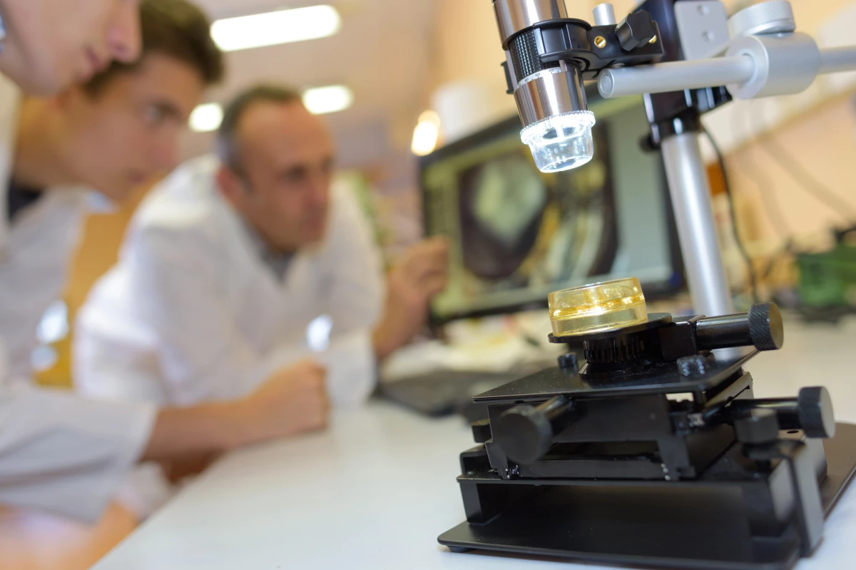
Prince David Okoro, the lead author of the study, emphasized the importance of this stability for conducting long-term research. “Mature brain cells are more reflective of real tissue function when investigating relevant diseases or traumas,” Okoro noted.
The construction of the scaffold involved a specially formulated liquid mixture of PEG, ethanol, and water, which results in a sponge-like structure filled with tiny pores. These pores facilitate the movement of oxygen and nutrients, ensuring that the stem cells thrive in their environment. Iman Noshadi, an associate professor of bioengineering at UCR, highlighted the potential of this material to mimic biological systems closely, enabling researchers to design tissue models with greater precision.
Future Applications and Aspirations
While the current scaffold measures just two millimeters across, the research team is focused on scaling up this technology. They have plans to explore its application in creating synthetic liver tissues as well. Their broader vision includes developing a network of lab-grown mini-organs that can communicate with one another, mimicking real biological systems.
“This interconnected system would allow us to observe how different tissues respond to the same treatment and how issues in one organ may impact another,” Noshadi explained. This approach represents a significant step toward a more integrated understanding of human biology and disease.
The layered fabrication technique used in this research not only enhances the functionality of the synthetic brain model but also opens new avenues for studying diseases, testing new drugs, and developing future treatments aimed at repairing or replacing damaged neural tissues. This breakthrough was documented in the journal Advanced Functional Materials, underscoring its potential impact on the field of bioengineering and medical research.